
How AI Filters Noise In Remote Cardiac Monitoring
See how Rhythm360 uses AI to filter ECG noise, reduce false alerts, and deliver clinically trusted cardiac monitoring data. Learn more.

Key Takeaways
- AI noise filtering in remote cardiac monitoring uses algorithms such as wavelet transforms, autoencoders, CNNs, and LSTMs to separate clinically meaningful cardiac events from motion artifact, EMG interference, baseline wander, power-line interference, and lead failure.
- A five-stage pipeline, including signal quality assessment, artifact classification, signal reconstruction, event detection, and false-alarm triage, reduces non-actionable alerts while preserving diagnostic accuracy.
- AI improves on traditional fixed filters by adapting to patient context and device type, while still requiring external validation, bias mitigation, and human oversight to manage subtle abnormalities and false negatives.
- Effective noise filtering improves clinical workflow, reduces alert fatigue, accelerates critical response times, and supports accurate CPT-billing documentation for remote monitoring programs.
- Rhythm360 applies vendor-neutral AI filtering across Medtronic, Boston Scientific, Abbott, Biotronik, and other devices to cut non-actionable alerts and support faster clinical response.
- Rhythm360’s implementation of this pipeline achieves greater than 99.9% data transmissibility through redundant data feeds, computer vision, and AI-powered extrapolation.
What Types Of Noise Does AI Filter In Cardiac Monitoring?
Each noise type in remote cardiac monitoring produces a distinct clinical consequence, and that mapping defines whether an AI filtering layer performs well.
Motion artifact arises from unstable skin-electrode contact during patient movement. Motion artifacts distort P-QRS-T morphology, interfere with R-peak detection and interval measurement, and increase both false-positive and false-negative AI outputs. Clinically, a patient walking to the bathroom can generate a transmission that resembles ventricular tachycardia.
EMG interference originates from skeletal muscle activity near electrodes. It superimposes high-frequency noise on the ECG baseline, mimics the irregular, rapid deflections of atrial fibrillation or ventricular flutter, and drives unnecessary alerts.
Baseline wander results from respiration, posture changes, or poor electrode adhesion. Bandpass filters with typical cutoffs between 0.5 and 150 Hz remove low-frequency baseline drift while preserving the diagnostically relevant ECG spectrum. When wander becomes severe, ST-segment shifts can be masked or fabricated, which can cause missed ischemia or false ischemia alerts.
Power-line interference at 50 or 60 Hz, depending on regional electrical standards, overlays a sinusoidal artifact that obscures fine waveform detail. Notch filters target this interference at 50 or 60 Hz, and fixed notch filters can also attenuate real signal components at those frequencies.
Lead failure produces abrupt signal dropout, flat-line segments, or high-amplitude noise bursts. Without accurate classification, lead failure is indistinguishable from asystole or ventricular fibrillation at the alert-triage layer, triggering emergency responses for a disconnected wire instead of a cardiac arrest. These noise types set the stage for understanding how AI filtering systems are designed to handle them.
How Does AI Filter Noise In Remote Cardiac Monitoring? Step By Step.
This five-stage pipeline describes how a well-designed AI filtering system processes a raw remote monitoring transmission from ingestion to actionable alert.
- Signal Quality Assessment. The pipeline begins by scoring incoming signal segments for usability. Hardware-informed gating using synchronized tri-axial IMU motion data and electrode impedance monitoring rejects motion- or contact-corrupted beats before software indices are applied. This approach prevents irrecoverably corrupted intervals from consuming downstream computation and produces a quality-labeled data stream in which only analyzable segments advance.
- Artifact Classification. Surviving segments are classified by noise type using CNNs for morphological pattern recognition and LSTMs for temporal sequence analysis. Hybrid CNN-LSTM models consistently outperform standalone CNNs or LSTMs by 3–8% on benchmark datasets, particularly for conditions involving both morphological abnormalities and rhythm disturbances. The output is a labeled artifact map that tells the reconstruction stage what to correct and what to preserve.
- Signal Reconstruction. Denoising autoencoders trained to reconstruct clean signals from noisy inputs learn to preserve diagnostic content while suppressing artifacts. Variational autoencoders are used for anomaly detection by learning the distribution of normal ECG patterns and identifying deviations. The result is a reconstructed signal that retains clinically important waveform features while removing noise-driven distortion.
- Event Detection. The cleaned signal is analyzed for arrhythmias, device-behavior anomalies, and physiologic changes. LSTM networks maintain internal memory states that can preserve information across hundreds of time steps, making them suitable for rhythm analysis such as atrial fibrillation detection and arrhythmia classification from long-term recordings. This stage produces a ranked list of detected events with associated confidence scores.
- False-Alarm Triage And Prioritization. Probability thresholding determines which events are surfaced to clinicians, which are archived as non-actionable, and which are flagged for human adjudication. Thresholding ensemble model predictions above 95% probability can reduce 91.9% of the classification workload while maintaining 100% sensitivity for AT/AF, with fewer than 0.1% errors. This stage produces a prioritized alert queue in which clinicians review only transmissions that warrant clinical attention.
Rhythm360 applies this AI-powered alert triage across a vendor-neutral, normalized data stream, ingesting signals from Medtronic, Boston Scientific, Abbott, Biotronik, and others to filter non-actionable noise and prioritize clinically significant events.
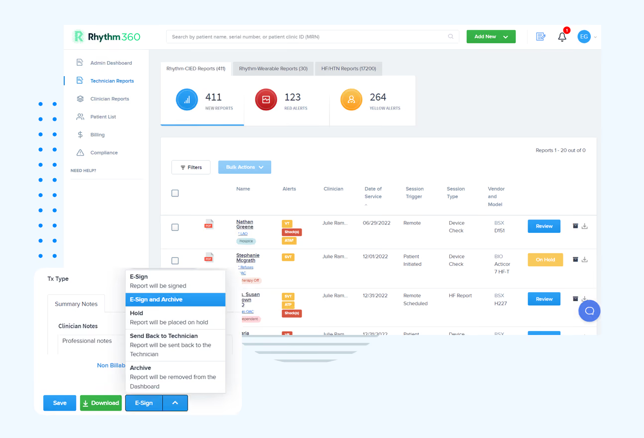
One of the most critical distinctions this pipeline must make is between a loose lead and a true arrhythmia, and AI handles that challenge with detailed beat-to-beat analysis.
Get A Noise-Filtering Workflow Review
How Does AI Distinguish A Loose Lead From A Real Arrhythmia?
The core mechanism relies on morphological and beat-to-beat analysis. A loose lead produces signal characteristics that differ systematically from true arrhythmia, including abrupt amplitude changes, absence of organized atrial or ventricular activation sequences, and noise bursts that do not follow physiologically plausible conduction patterns.
A 2026 multicenter study published in Sensors by van Krimpen et al. trained a 1D residual neural network ensemble on atrial high-rate episodes from Biotronik pacemakers and ICDs across five hospitals, achieving precision of 97.2–99.4% for true AT/AF, 93.1–97.7% for noise, and 75.4–94.4% for far-field oversensing, while maintaining 98.9–99.3% sensitivity for AT/AF. The model used Grad-CAM++ saliency maps to confirm focus on clinically relevant signal patterns, such as high-frequency atrial noise for noise episodes, morphologically similar ventricular activations for far-field oversensing, and absence of consistent atrioventricular conduction for atrial fibrillation.
The practical implication is that beat-to-beat morphological consistency, assessed across a sliding window rather than a single beat, serves as the primary discriminator. A true arrhythmia maintains internal electrophysiological logic even when its rate is abnormal. A loose lead does not.
A cross-manufacturer analysis presented at the 2026 EHRA Congress found that 46.8% of device-labeled pause episodes were false positives caused by R-wave undersensing. This finding illustrates how device-level artifact classification and cloud-based AI filtering address different layers of the same problem.
AI Noise Filtering vs. Traditional Filters In Cardiac Monitoring
While AI can distinguish complex scenarios like loose leads from arrhythmias, it differs in fundamental ways from the traditional filters long used in cardiac monitoring.
Traditional fixed filters, including bandpass, notch, and wavelet-based designs, operate on predetermined frequency cutoffs and transform parameters. They perform reliably when noise characteristics remain stable and predictable, and they cannot adapt when a patient’s physiology or environment changes.
Conventional rule-based ECG interpretation systems are often limited by noisy recordings, signal artifacts, rigid diagnostic thresholds, and poor generalizability across diverse patient populations. A fixed bandpass filter set for a resting adult will misclassify artifacts generated by an ambulatory patient with a high resting heart rate because the noise and signal spectra overlap.
Adaptive AI models learn the statistical structure of both signal and noise from large annotated datasets and update their internal representations as new data arrives. Denoising autoencoders trained to reconstruct clean signals from noisy inputs learn to preserve diagnostic content while suppressing artifacts. Fixed filters cannot replicate this because they have no representation of what “diagnostic content” means.
The practical advantage is context-sensitivity. An adaptive model can recognize that a high-amplitude deflection in a pacemaker patient is a pacing spike rather than a QRS complex and adjust its reconstruction accordingly. A fixed filter applies the same transformation regardless of device type or patient context.
Can AI Filter Motion Artifacts In Wearable ECG?
Wearable and implantable data streams have fundamentally different noise profiles, and they require different filtering approaches.
Implantable devices acquire intracardiac or subcutaneous electrograms in a mechanically stable environment. The dominant noise sources are far-field oversensing, electromagnetic interference, and lead-related artifacts. Signal amplitude is high relative to noise, and the acquisition geometry remains fixed.
Wearable ECG is acquired in uncontrolled ambulatory environments. Wearable ECG is highly vulnerable to baseline wander from respiration or posture change, electrode-motion artifacts from unstable skin-electrode contact, and muscle artifacts from skeletal muscle activity. Single- or few-lead acquisition provides less spatial information than 12-lead ECG, which constrains some diagnostic tasks.
Advanced AI architectures integrate concurrent kinematic data from triaxial accelerometers to identify and discard periods of excessive physical exertion, restricting rhythm analysis exclusively to intervals of high signal fidelity and thereby minimizing false alerts. This cross-referencing of motion sensor data with ECG signal does not apply to implantable devices, which lack external motion references.
Wearable ECG is more susceptible to motion artifact and noise than traditional 12-lead ECG because it is used in ambulatory, real-world settings. This limitation means wearable AI filtering must be evaluated separately from implantable device filtering, using metrics appropriate to each context.
What Are The Limitations Of AI Noise Filtering In Cardiac Monitoring?
AI noise filtering carries documented risks that clinicians and program managers must understand before they trust a filtered alert stream with patient safety.
Suppression of clinically subtle abnormalities. A 2026 study published in BioData Mining reported that a diffusion-based reconstruction model struggled to reconstruct F waves in atrial fibrillation patients, demonstrating that noise filtering or signal reconstruction can suppress clinically subtle abnormalities. The same study noted that “even minor distortions can lead to misinterpretations in medical assessments” and that conventional generative metrics like RMSE do not capture these clinical losses.
Lack of external validation. A 2026 systematic review published in Discover Artificial Intelligence found that external validation was lacking in 72% of cases. Cross-dataset validation studies show average performance degradation of 8–15% when models are tested on data from different sources. A model that performs at 98% accuracy on its development dataset may perform materially worse on a different patient population, device manufacturer, or clinical setting.
Dataset bias. Seventy-eight percent of public ECG datasets originate from North America or Europe, ethnicity data is reported in fewer than 10% of datasets, and average female representation across datasets is 41.5%. A model trained on this distribution may perform differently in a diverse urban practice.
Black-box opacity. Many deep learning ECG models remain inherently black-box systems, making it difficult for clinicians to understand the reasoning behind individual predictions. Without interpretability, a clinician cannot determine whether a filtered-out transmission was correctly archived or incorrectly suppressed.
Clinicians should demand several safeguards from any AI filtering vendor. These include published external validation on a population comparable to their own patient panel, reported false-negative impact alongside false-positive reduction, evidence that low-amplitude diagnostically important features are preserved after reconstruction, and a human-in-the-loop review layer for low-confidence predictions. Sensitivity and specificity are only part of the picture; predictive values, calibration, false-positive burden, and performance at the intended decision threshold may matter more at the bedside.
How Does AI Handle Pacemaker Lead Failure Alerts?
Pacemaker lead failure produces a characteristic signal signature that includes loss of pacing capture, impedance changes, or abrupt signal dropout. The clinical consequence of misclassifying lead failure as a true arrhythmia, or vice versa, is significant. A missed lead failure can result in failure to pace in a pacemaker-dependent patient. A false lead-failure alert triggers an unnecessary urgent evaluation.
AI distinguishes true lead failure from noise by analyzing multiple concurrent signal features, including pacing spike morphology, capture confirmation (the presence of a QRS complex following each pacing spike), impedance trend data, and beat-to-beat consistency. Lead failure typically produces an abrupt change in all of these parameters simultaneously. Noise artifacts, by contrast, affect signal amplitude without altering pacing capture patterns.
Even in AI-equipped devices, 32.9% of episodes were non-actionable and another 30.6% were indeterminate in a cross-manufacturer analysis of 2,659 rhythm episodes from 1,710 patients presented at the 2026 EHRA Congress. This pattern underscores that device-level AI and cloud-based AI filtering address complementary but distinct layers of the alert pipeline. Cloud-based AI applied to normalized, cross-manufacturer data streams can apply consistent classification logic that individual device algorithms, each trained on proprietary data, cannot.
Talk With Our Clinical Workflow Team
How Filtered Data Feeds Alert Triage And CPT-Billable Documentation
Each stage of the AI filtering pipeline has a direct downstream effect on clinical workflow and billing documentation.
Signal quality assessment determines which transmissions contain analyzable data. Transmissions that fail quality gating are flagged for technical follow-up rather than clinical review, which reduces the volume of transmissions that reach a clinician’s queue. Seventy-three percent of CIED alerts and 90% of scheduled transmissions are non-actionable, with only 7% of alerts leading to in-office follow-up. This distribution means the majority of clinical review time is currently spent on transmissions that do not require it.
Artifact classification and reconstruction determine whether a transmission contains a true event or a noise artifact. Accurate classification at this stage prevents both missed events and unnecessary urgent responses. Combining AI software with human review and alert-based monitoring allows high-performing monitoring programs to archive over 50% of non-actionable transmissions without sacrificing clinical accuracy.
False-alarm triage and prioritization determine the order in which clinicians review transmissions. A prioritized queue enables faster response to critical events and generates the documented review record required for CPT billing.
The documented review record that triage generates is what supports CPT billing. Remote monitoring CPT codes are tied to specific device types and billing cycles. Pacemakers and ICDs use a 90-day cycle: 93294 (pacemaker professional), 93295 (ICD professional), and 93296 (pacemaker/ICD technical). Physiologic monitors and loop recorders use a 30-day cycle: 93297 covers implantable cardiovascular physiologic monitors such as CardioMEMS, and 93298 covers subcutaneous cardiac rhythm monitors and implantable loop recorders. Each of the 30-day codes is billable once per 30 days and can be billed global, with a -26 professional modifier, or with a -TC technical modifier. Rhythm360 automatically tracks device-specific CPT pairings and generates the documentation required to support each code, helping practices improve billing accuracy and revenue outcomes.
Frequently Asked Questions
Can AI Reliably Read An EKG?
AI can classify ECG findings with reported accuracy commonly ranging from 90% to 99% on benchmark datasets, with sensitivity and specificity frequently exceeding 90% in controlled validation studies. Performance degrades when models are applied to patient populations, device types, or clinical settings that differ from their training data, a phenomenon known as dataset shift. AI ECG interpretation is most reliable as a decision-support tool reviewed by a qualified clinician, rather than as a standalone diagnostic system.
How Is AI Used In Remote Patient Monitoring?
In remote cardiac monitoring, AI is applied across the full transmission pipeline. It scores signal quality, classifies artifact types, reconstructs clean signals, detects arrhythmias and device anomalies, and prioritizes alerts for clinical review. AI also supports automated report generation and CPT-billable documentation. The most effective implementations combine AI-powered triage with human clinical review for low-confidence or ambiguous findings, which preserves sensitivity for true events while reducing the volume of non-actionable transmissions that reach clinicians.
What Is Signal Quality Assessment In ECG AI?
Signal quality assessment is the first stage of the AI filtering pipeline and evaluates incoming ECG segments for usability before any diagnostic analysis is applied. Assessment methods include hardware-informed gating using electrode impedance and motion sensor data, frequency-domain signal-to-noise ratio estimation, morphological consistency checks against adaptive beat templates, and physiological plausibility constraints on RR intervals. Segments that fail quality thresholds are labeled and excluded from downstream analysis, which prevents corrupted data from generating false alerts or entering model training pipelines.
What Is Autoencoder ECG Noise Removal?
An autoencoder is a neural network architecture trained to compress an input signal into a lower-dimensional representation and then reconstruct it. A denoising autoencoder is trained specifically on pairs of noisy and clean ECG signals, learning to reconstruct the clean version from the noisy input. The reconstruction process suppresses noise components that are not part of the learned signal distribution while preserving diagnostically important waveform features. As noted in the limitations section, reconstruction models can smooth or suppress low-amplitude features such as F waves in atrial fibrillation that are clinically significant but statistically rare in training data.
Explore Rhythm360 For Your Program
Rhythm360: Vendor-Neutral AI Filtering Built For Clinical Trust
Electrophysiologists, device technicians, and RPM program managers face a compounding problem. Transmission volumes exceed what any team can manually review. Alert streams mix genuine emergencies with noise artifacts. Device data is siloed across manufacturer portals that do not communicate with each other. The result is alert fatigue, missed events, and billing documentation that falls short of CPT requirements.
Rhythm360 addresses this problem at the data layer. As mentioned earlier, Rhythm360’s vendor-neutral approach ingests and normalizes signals from all major CIED manufacturers into a single platform and applies consistent AI-powered alert triage logic across the entire patient population regardless of device brand. As noted above, the platform achieves greater than 99.9% data transmissibility through redundant data feeds, computer vision, and AI-powered extrapolation, which helps ensure that the filtered alert stream reflects the most complete and accurate view of each patient’s status.
Practices that implement Rhythm360 capture previously lost revenue through stronger CPT code documentation and benefit from bi-directional EHR integration with Epic, Cerner, Athenahealth, eClinicalWorks, and others. This integration eliminates manual transcription and supports the auditable documentation that remote monitoring billing requires.
Read Next
- AI in Cardiac Telemetry: Reduce False Alarms & Unify Data
- AI Alerts Transform Heart Failure Monitoring & Care
- AI Triage Heart Failure Alerts: Cut Alert Fatigue by 80%
- AI Cardiac Monitoring Remote Patient Care: A Practice Guide
- AI Workflow Automation in Cardiac RPM: A Practical Guide for Cardiology Practices
.avif)